Home
/ Edge Of Paper Under Microscope : Scanning Electron Microscopy A Sheet Of Notebook Paper Cut With A Knife Torn, Why should a microscope slide and coverslip be held by their edges?
Edge Of Paper Under Microscope : Scanning Electron Microscopy A Sheet Of Notebook Paper Cut With A Knife Torn, Why should a microscope slide and coverslip be held by their edges?
Edge Of Paper Under Microscope : Scanning Electron Microscopy A Sheet Of Notebook Paper Cut With A Knife Torn, Why should a microscope slide and coverslip be held by their edges?. Using a dropper, place one or two drops of iodine solution along one edge of the cover slip. You can place each torn paper in separate (marked/labeled) petri dishes so that you keep a record of each paper you observed. As a company, foldscope instruments inc's mission is to break down the price barrier between people & the curiosity and excitement of. The paper towel will draw the water out from under the coverslip, and the cohesion of water will draw the stain under the slide. Fiber identification generally involves taking samples from the artifact and viewing them at 100 times or greater magnification to study the fiber morphology.
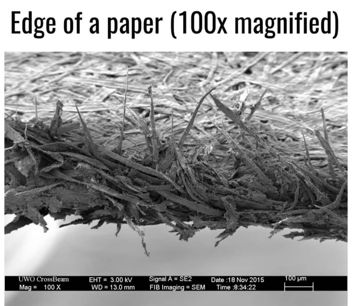
So it doesn't fall or get knocked over. But to add, this is a scanning electron microscope (sem) image which usually isn't as high mag as a good transmission electron microscope (tem) that you might be thinking of. Cheap toilet paper (single ply) iodine solution or water. Wood pulp, on the other hand, has no characteristic features like that of a fiber structure. Capillary action will pull the dye across the slide to stain the specimen.
Edge Of Paper Magnified 9gag from images-cdn.9gag.com As soon as the stain has covered the area containing the specimen, you are finished. The paper towel will draw the water out from under the coverslip, and the cohesion of water will draw the stain under the slide. The paper is lens cleaning paper that i use to clean my microscope slides, and, of course, lenses, but reuse them like this when dried to guard against spilling alcohol, etc. Why should a specimen being viewed under a microscope should be thin? Pick up the coverslip by its edges. Wood pulp, on the other hand, has no characteristic features like that of a fiber structure. Why is it a good idea to place the microscope at least ten centimeters from the edge of the table? Click card to see definition 👆.
Tear a tiny piece of toilet paper off the sample and mount it in water or iodine solution.
Pick up the coverslip by its edges. Capillary action will pull the dye across the slide to stain the specimen. This extremely thin edge curls a bit since it is thin and fragile. Add a small drop of stain to an edge of the coverslip. Why is it a good idea to place the microscope at least ten centimeters from the edge of the table? Prepare a wet mount or dry mount with a coverslip. Note the thin curl of metal at the edge of the blade. Place the flat edge of a piece of paper towel on the opposite side of the coverslip. The amazing thing about paper is how strong it is. Using a dropper, place one or two drops of iodine solution along one edge of the cover slip. In contrast, the light has to pass through the specimen to form the image under a compound microscope. As soon as the stain has covered the area containing the specimen, you are finished. You can place each torn paper in separate (marked/labeled) petri dishes so that you keep a record of each paper you observed.
The actual sharpening of the cutting edge begins here. Some sand is made up largely of. The amazing thing about paper is how strong it is. Place a paper towel sheet against one edge of the cover slip. Why should a specimen being viewed under a microscope should be thin?
Fascinating Images Of Everyday Objects Under A Microscope Reader S Digest from www.rd.com If you put it too close, it can fall off and break. In contrast, the light has to pass through the specimen to form the image under a compound microscope. Under normal conditions, resolution is increased by decreasing the wavelength of the light source; Stains are often employed to accentuate features and to determine pulping processes. The edge of a piece of paper at 100x under electron microscope. The edge itself is being reduced down to a foil thin point. Place the flat edge of a piece of paper towel on the opposite side of the coverslip. The housefly under a stereo microscope.
The paper is lens cleaning paper that i use to clean my microscope slides, and, of course, lenses, but reuse them like this when dried to guard against spilling alcohol, etc.
If we also examine a paper made of cotton rag, it will have characteristics of cotton fibers. Cheap toilet paper (single ply) iodine solution or water. When drawn over food, it glides along because there is so little friction. Set the towel against the slip's edge without disturbing the material under the cover slip. Once the slides are prepared, have students use paper and pencils to create a chart with two columns. The edge of a piece of paper at 100x under electron microscope. Some sand is made up largely of. Stand the coverslip on its edge next to the drop of water. Note the thin curl of metal at the edge of the blade. It is like some slices of multiple slices of wood mixed. Under normal conditions, resolution is increased by decreasing the wavelength of the light source; They should label the first column euglena, and the second column paramecium. have students place the euglena slide on the microscope stage. This extremely thin edge curls a bit since it is thin and fragile.
Capillary action will pull the dye across the slide to stain the specimen. They should label the first column euglena, and the second column paramecium. have students place the euglena slide on the microscope stage. Focus on the torn edge of the paper and observe the long sclerenchyma fibres. Place the edge of a tissue or paper towel on the opposite edge of the coverslip. Wood pulp, on the other hand, has no characteristic features like that of a fiber structure.
Edge Detection In Scanning Electron Microscope Sem Images Using Various Algorithms Semantic Scholar from d3i71xaburhd42.cloudfront.net It is like some slices of multiple slices of wood mixed. Why should a microscope slide and coverslip be held by their edges? Position lens #1 in the hole in the middle of the microscope tube so that the raised white edge of the lens holder is closer to your eye. Add a small drop of stain to an edge of the coverslip. The edge itself is being reduced down to a foil thin point. As a company, foldscope instruments inc's mission is to break down the price barrier between people & the curiosity and excitement of. This extremely thin edge curls a bit since it is thin and fragile. But to add, this is a scanning electron microscope (sem) image which usually isn't as high mag as a good transmission electron microscope (tem) that you might be thinking of.
Prepare a wet mount or dry mount with a coverslip.
The ink looks solid normally, but under a microscope it looks broken up and has ink smudges. The absorptive paper towel will draw some of the water out from under the cover slip, and pull the staining agent under the cover slip and onto the specimen. Kostenlose stornierung bis zu 24 stunden vor ihrer aktivität. Cheap toilet paper (single ply) iodine solution or water. Why should a specimen being viewed under a microscope should be thin? Click again to see term 👆. It is like some slices of multiple slices of wood mixed. Why is it a good idea to place the microscope at least ten centimeters from the edge of the table? Do not touch the surface of the coverslip. Tap card to see definition 👆. Newspaper print under the microscope you will notice that if you take a close look at your favorite printed newspaper you will see that the black and white photos are actually made from thousands of tiny dots all spaced out in ways that make the blacks darker and the greys lighter. The housefly under a stereo microscope. Fiber identification generally involves taking samples from the artifact and viewing them at 100 times or greater magnification to study the fiber morphology.
Click card to see definition 👆 paper under microscope. Tear a tiny piece of toilet paper off the sample and mount it in water or iodine solution.
